The Accelerating Impact of Smoking on Brain Aging
For a long time, smoking has been associated with various health issues, including lung cancer and heart disease. Recent research has added another significant concern: accelerated brain aging. Studies indicate that smokers have up to a 20% higher risk of cognitive decline compared to non-smokers, underscoring the severe impact smoking has on brain health. Understanding how smoking impacts brain health and contributes to accelerated brain aging is crucial for public health awareness and smoking cessation efforts.
Reduction in Brain Volume
Studies have consistently shown that smokers tend to have a reduced brain volume compared to non-smokers. This reduction is especially pronounced in regions like the hippocampus, which is essential for learning and memory. Brain volume loss in these regions can lead to significant cognitive impairments, affecting everyday activities and overall quality of life.
Detection Methods:
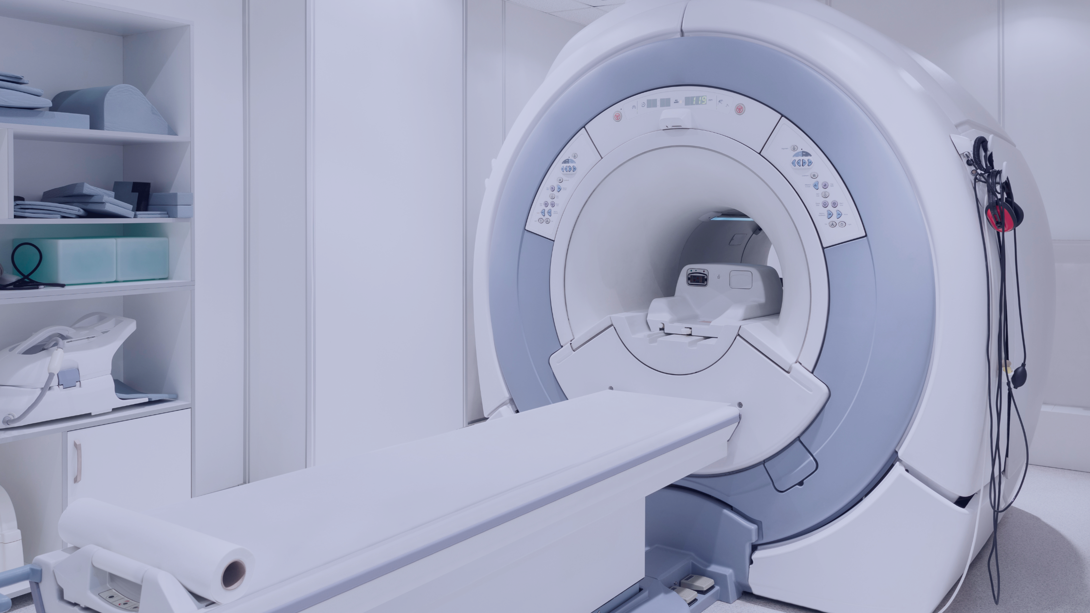
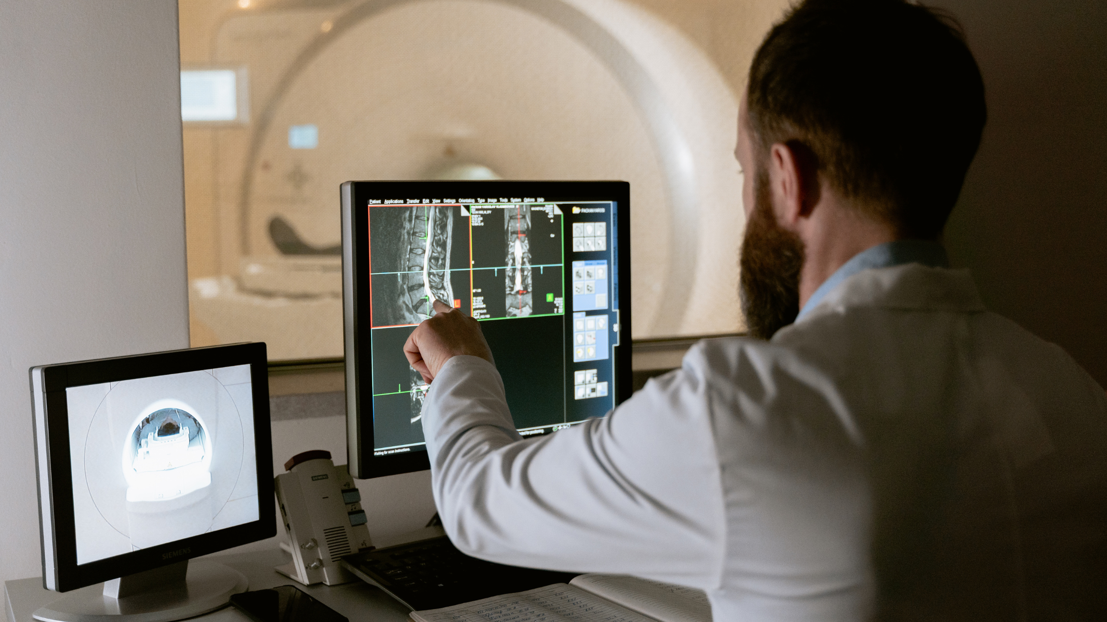
Sophisticated diagnostic methods, including Magnetic Resonance Imaging (MRI) and Computed Tomography (CT), scans are used to detect this reduction in brain volume. MRI, in particular, provides detailed images of brain structures, allowing researchers and clinicians to accurately measure the volume of different brain regions.
White Matter Integrity
White matter, which is responsible for communication between different brain regions, is also adversely affected by smoking. Damage to white matter can lead to cognitive decline and impaired brain function. The integrity of white matter is essential for efficient brain connectivity and overall cognitive health, and its deterioration can have profound consequences on mental processes and coordination.
Detection Methods:
Diffusion Tensor Imaging (DTI), a type of MRI, is specifically used to assess the integrity of white matter. DTI measures the diffusion of water molecules along white matter tracts, providing insights into the condition and connectivity of these crucial pathways.
Cerebral Blood Flow
Nicotine and other chemicals in cigarettes adversely affect cerebral blood flow. Reduced blood flow means less oxygen and fewer nutrients are delivered to the brain, impairing brain function and leading to accelerated aging. Efficient cerebral blood flow is crucial for maintaining the health and functionality of brain tissues, and any disruption can have immediate and long-term adverse effects.
Detection Methods:
(PET) and MRI (fMRI) are commonly used to measure cerebral blood flow. These imaging techniques can assess how well blood flows to various brain parts, providing valuable information about potential impairments.
Oxidative Stress and Inflammation
Smoking increases oxidative stress and inflammation throughout the body, including the brain. These processes can damage brain cells and accelerate the aging process. When there is a disruption in the balance between antioxidants and free radicals within the body, oxidative stress arises, harming cells. Additionally, prolonged inflammation in the brain can worsen damage to neurons and play a role in the advancement of neurodegenerative conditions.
Detection Methods:
Blood tests can measure biomarkers of oxidative stress and inflammation. Additionally, PET scans can detect neuroinflammation in the brain, visually representing these harmful processes.
Cognitive Decline
Research has shown that smokers are at a higher risk for cognitive decline and dementia. The damage caused by smoking can lead to impairments in memory, attention, and executive functions. Cognitive decline can severely impact daily living, reducing the ability to perform routine tasks and decreasing overall life satisfaction.
Detection Methods:
Neuropsychological tests are employed to assess cognitive functions. These tests evaluate memory, attention, problem-solving, and other mental abilities. Furthermore, MRI and PET scans help detect anatomical and physiological alterations in the brain linked to cognitive deterioration.
Increased Risk of Neurodegenerative Diseases:
Smoking has been linked to an increased risk of neurodegenerative diseases such as Alzheimer’s disease and Parkinson’s disease. These conditions are associated with aging, and smoking can exacerbate their onset and progression. The toxins in cigarette smoke can accelerate the pathological processes underlying these diseases, leading to earlier and more severe manifestations.
Detection Methods:
MRI and PET imaging techniques play a crucial role in the diagnosis and ongoing observation of the progression of neurodegenerative disorders. These imaging techniques help visualize brain atrophy, abnormal protein deposits, and other pathological changes.
Genetic Factors
Genetic predispositions can increase the vulnerability of some people to the negative impacts of smoking on brain health. For example, variations in the CHRNA5 gene, which is involved in nicotine dependence, can influence the degree to which smoking accelerates brain aging. Understanding genetic predispositions can help tailor more effective smoking cessation programs and preventative measures for those at higher risk.
Conclusion
The evidence indicates that smoking accelerates brain aging through a variety of mechanisms, including brain volume reduction, white matter damage, impaired blood flow, oxidative stress, and increased risk of cognitive decline and neurodegenerative diseases. Quitting smoking can help mitigate some of these effects, highlighting the importance of smoking cessation programs and interventions for brain health.
Public health campaigns must continue to emphasize not only the general health risks of smoking but also its profound impact on brain health. As more people become aware of these risks, the hope is that smoking rates will decline, leading to healthier, longer lives and sharper minds in the aging population.